|
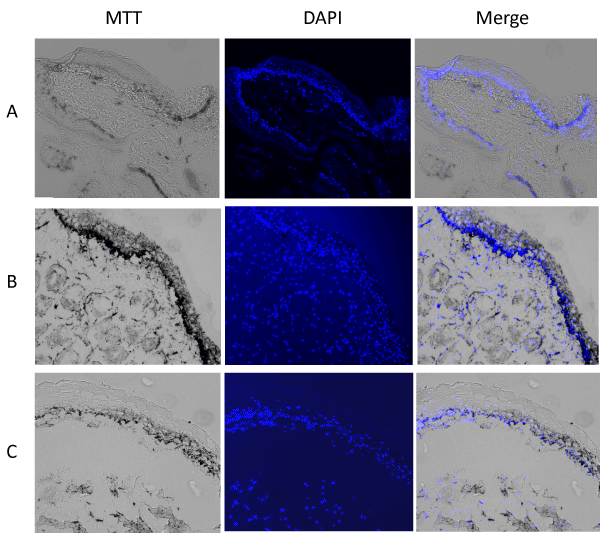
| Figure 4: MTT deposition in DMBA-initiated HCP mucosa. HCPs were excised from (A) sentinel control hamsters; (B) DMBA (4 wks) followed by 2% methylcellulose 24 h; (C) DMBA (4 wks) followed by 2% D003 extract 24 h. The HCPs were incubated with MTT, processed into 10 μm sections (left panel), and then co-stained with DAPI (middle panel) as described in Materials and Methods. MTT deposition shows as dark and black color (left and right panel). Nuclei were stained using DAPI and appear blue (middle and right panel). |