 |
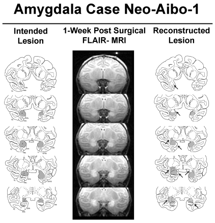
| Figure 1: Intended amygdala damage is shown in gray on coronal sections through the amygdala of an infant macaque brain atlas in the left column. Location of hypersignals shown on FLAIR MR coronal images is given at several matched anterior-posterior levels through the amygdala in case Neo-Aibo-1 (middle column). Edema caused by cell death appears white within and around the amygdala. The estimated reconstructed lesion extent is shown in the right column. Arrows point to areas of unintended damage or sparing. Abbreviations: ls – lateral sulcus; sts – superior temporal sulcus; ots – occipital temporal sulcus; ERh – entorhinal cortex; PRh – perirhinal cotex; TE, temporal cortical area and TH/TF – cytoarchitectonic fields of the parahippocampal gyrus. |