|
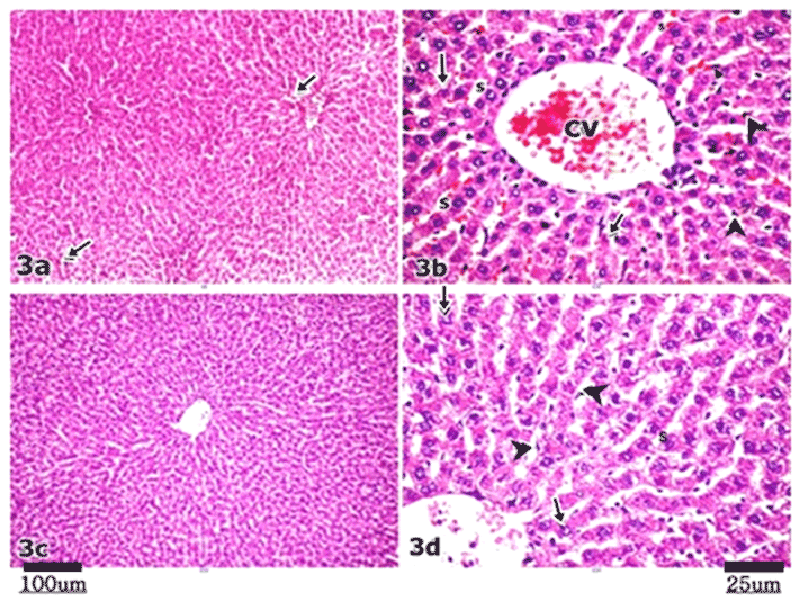
| Figure 3: Micrograph of TOTM group (III). (a): shows preserved hepatic lobular architecture with dilatation and congestion of the blood vessels (arrow), Subgroup IIIA. (b): Higher magnification (a) shows congested central vein (cv) and blood sinusoids (s) with prominent kupffer cells (arrowhead). Hepatocytes appear shrunken (arrow). (c): shows hepatic lobule with apparent normal lobular architecture, Subgroup IIIB (d): Higher magnification (c) shows plates of normal hepatocytes with rounded vesicular nuclei (arrow) and slightly dilated blood sinusoids (arrowhead). |