|
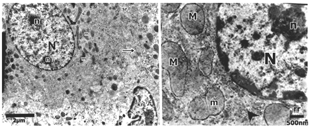
| Figure 6: An electron micrograph of hepatocyte in the control group liver. (a): shows hepatocyte with euchromatic nucleus (N) with two prominent nucleoli (n). The cytoplasm shows many mitochondria cisternae of rER (rr) meshwork of smooth endoplasmic reticulum (sr) tubules were distributed in the cytoplasm. Intercellular junction and kupffer cell with indented nucleus were also observed. (b): shows euchromatic nucleus (N) with prominent nucleolus (n). The cytoplasm contains mitochondria (M); one of them appears in binary fission (m). Cisternae of rough endoplasmic reticulum (rr) and glycogen granules (arrowhead) are also seen. |