|
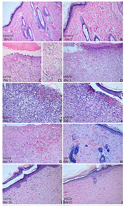
| Figure 2: HE stained sections of post-wounding recovering tissue. Photomicrographs of mice skin and scar tissue with or without treatment, stained with HE technique. Full-thickness excisions from the wounding site topically treated with pM2b (50 μl, 230 nM) or saline were subjected to histological analysis. Fragment of control skin with ortho keratinized epidermis. Dermis is composed by dense connective tissue, intermingled by skin appendages. Deep aspects of the specimens show striated muscle bundles and adipose tissue. |