|
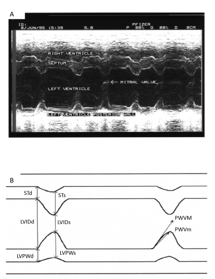
| Figure 6: M mode recording in dogs. (A) example of tracing recorded from a long axis section in two-dimensional mode. The guidance line is positioned at the tip of the mitral valve. The movements of the septum and free wall of the left ventricle are followed over time. (B) Schematic representation of measured parameters. |