|
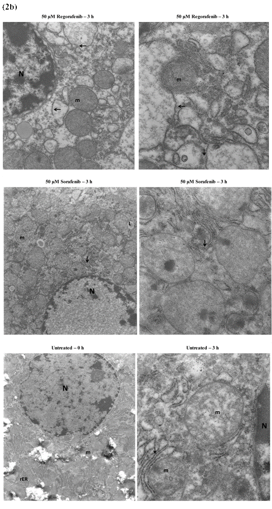
| Figure 2B: Untreated tissue at 0 h, showing a hepatocyte with preserved architecture; magnification =7,000. The white areas represented an artifact from specimen collection or preparation. Untreated tissue at 3 h, showing a hepatocyte with relatively preserved cellular architecture. Note hepatocyte nucleus (N), mitochondria (m) with minimal swelling, relatively preserved rER (arrow) with attached ribosomes; magnification =27,500. Tissue treated with 50 μM sorafenib at 3 h, showing a hepatocyte with swollen mitochondria and relatively preserved rER. The focal minimal detachment of ribosomes (arrows) from rER was similar to untreated liver at 3 h; magnification =27,500.Tissue treated with 50 μM regorafenib at 3 h,showing a hepatocyte with mildly distended mitochondria (m), significant disruption of the rER and focal detachments of the ribosomes (arrows); magnification =14,000. |