|
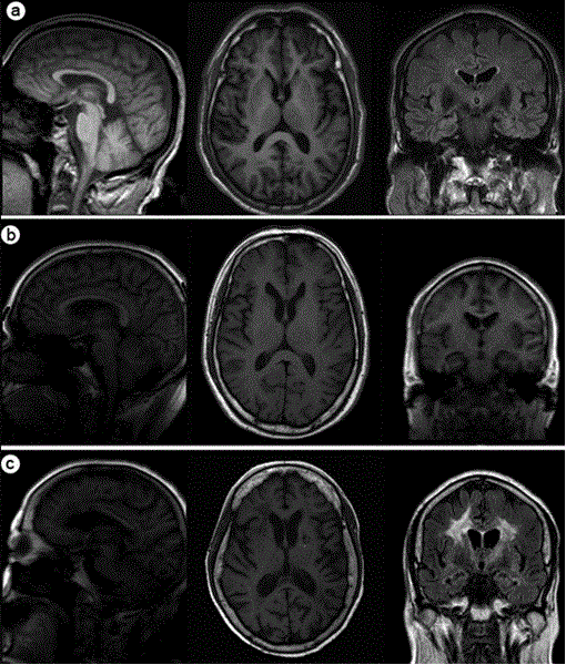
| Figure 2: Brain-volume deficits in alcoholism and its sequelae. Sagital (left column), axial (middle column) and coronal (right column) brain T1-weighted MRI are shown. (a) a 65-year-old healthy control male, ( b) a 64-year-old man with alcoholism, and c. a 50-year-old man with alcoholism. Enlargement of the ventricles (b, c) can be observed compared with the healthy control (a), which indicating shrinkage of the surrounding tissue. Leukoaralosis around the ventricles can be observed in picture c The abnormal signal around the ventricles (c) was leukoaralosis. |