|
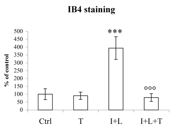
| Figure 3: Effects of THC on microglial activation. Aggregate cultures remained either untreated [Ctrl] or were treated with THC (1 μM) [T]; IFN-γ (50 U/ml) plus LPS (5 μg/ml) [I+L]; or treated with THC simultaneously with the inflammatory agents [I+L+T]. Cultures were harvested 48 hours after the last treatment with the inflammatory agents. The IB4- labelled microglial cells were quantified by measuring 20 aggregate sections per treatment and expressing the stained area as percent of untreated control cultures. The Figure shows representative data from one experiment. Results were statistically evaluated for significance by one-way ANOVA test followed by the Tukey post-test. (***P<0.001 compared with untreated control cultures; °°°P<0.001 compared with to cultures treated with the inflammatory agents). |