 |
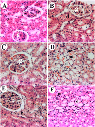
| Figure 2: Kidney paraffin sections stained by haematoxylin and eosin (H&E) for histopathological changes. Control group [A] showing intact histological structure of glomeruli (g) and renal tubules (t) (x 80), Cyr-treated group [B] showing vacuolization and swelling in the endothelium of glomerular tuft (g) with swelling in the lining epithelium of tubules (t) (x 80), CPF-treated group [C &D] showing congestion in the tuft of the glomeruli (g) (x 80) and inflammatory cells infiltration (m) in between the degenerated tubules (d), Cyr+CPF-treated group [E&F] showing swelling and vacuolization in the endothelial cells lining the tuft of the glomeruli (g) with fibrosis (f) and hyalinosis (h) between the tubules in focal manner and inflammatory cells infiltration (m) and few fibroblastic cells proliferation (arrow) in between the degenerated tubules (d) in focal manner at the corticomedullary portion (x 80). |