|
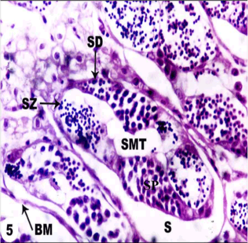
| Figure 5: Photomicrograph of T.S. of testes of O. niloticus of LM April showing the reduction in the amount spermatocytes (SP), spermatids (SD) & the spermatozoa (SZ) cells in the seminiferous tubules (SMT), obvious separation (S) of the boundary membrane of seminiferous tubules (BM) and presence of many vacuoles in interstitial cells and between spermatogenic cells (V). (X 400). |