|
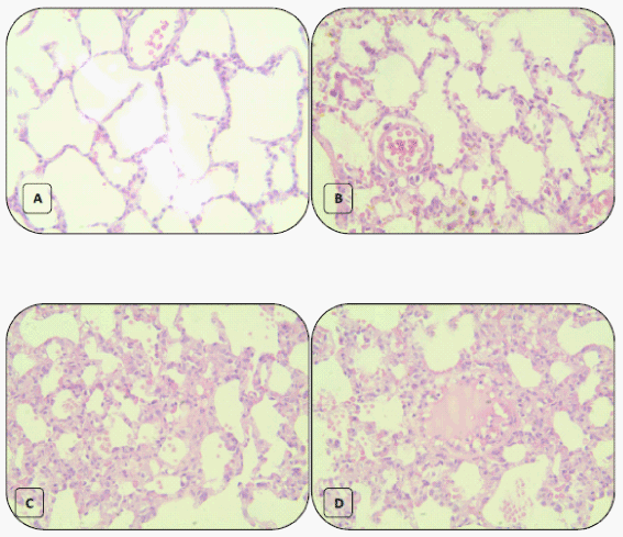
| Figure 10: Histopathological changes in lung tissue from (a) control and Ajwa extract groups showing normal alveolar cappilaries, alveolai and alveolar walls. (b) Combined Ajwa extract-lead intoxicated group showing mild alveolar cappilary congestion, thickening of alveolar walls, mild alveolar Inflammatory cellular infiltrate. (c and d) Lead intoxicated group without treatment showed marked alveolar capillaries congestion, alveolar wall thickening, pink coagulum in the alveoli, marked alveolar inflammatory cellular infiltrates, moderate alveolar walls destruction and emphysematous changes were observed (H and E stain, 40x). |