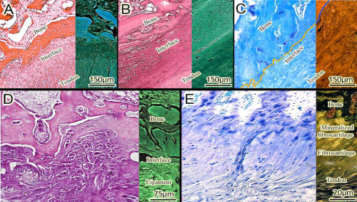
The insertion of the native tendon is characterized in four layers: tendon, fibrocartilage, mineralized fibrocartilage, and bone (A to E). The collagen fibers of the tendon extend into both the fibrocartilage and the mineralized layer (E). The tendon-bone healing process occurs as follows: First, fibrovascular interface tissue forms between the graft and bone, and progressive mineralization of the interface tissue occurs with subsequent bone in growth into the outer tendon and incorporation of the tendon graft into the surrounding bone. Sharpey’s fibers are made up of type I collagen and connect the periosteum to the bone. Progressive reestablishment of the continuity of collagen fibers between the tendon and the bone results in re-establishment of a tendoosseous junction. Tendoosseous junction is the interface zone (A to D). A and B stained with H and E. C to E stained with toluidine blue.