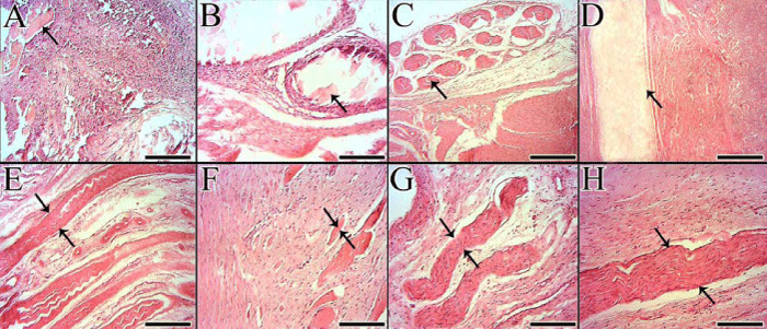
Implantation of various prosthetic implants causes different host behavior based on the nature of the implant. Implantation of the highly antigenic implants cause rapid absorption (A and B) which is described as one of the host rejection mechanisms. Vigorous inflammatory reaction occurs after implantation of the antigenic graft (A) and the inflammatory cells surround the scaffold remnants to completely lyse them at a short period of time. Arrows in A and B show the scaffold remnants. The host can reject the implanted graft by another mechanism which is called encapsulation (C). In fact, the body fails to lyse all parts of the graft, thus the fibrous connective tissue surrounds the graft (C, arrow). If the implant would not be rejected by the host then it can collaborate at different stages of tendon and ligament healing and regeneration by different mechanisms (D to H). D to F is progressive degradation mechanism in which some part of the implant have been absorbed and the new tissue filled the free spaces in the graft and the remnant parts (arrow) are not surrounded by the inflammation and are not encapsulated. The remnant parts are progressively degraded at later stages and the new tissue fills their free spaces (D). The absorption rate in this mechanism is high but is lower than the acute degradation mechanism which we showed in A and B. The final mechanism is adoption and adaptation of the graft (G and H). Some remnants of the graft (arrows) are not rejected by the host, also are not slowly degraded but also infiltrated by the host fibroblasts and accepted as part of the new tendon or ligament. Color staining: H & E. Scale bar for A, C, D, and E: 125 μm and for B, F, G and H: 50 μm.