 |
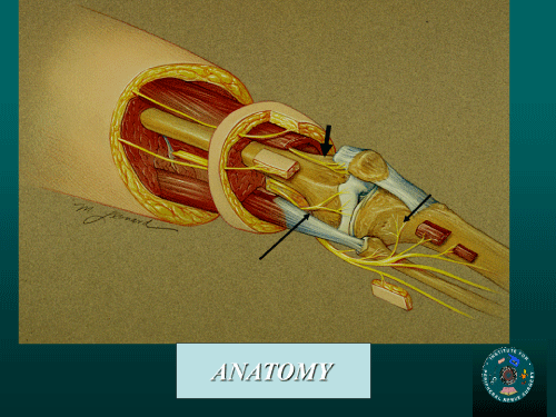
| Figure 2: Lateral view of the innervation of the knee. The skin in this area represents the terminal branches of the lateral femoral cutaneous nerve. The lateral joint structures are innervated by a branch of the sciatic nerve that comes from the popliteal fossa, goes deep to the biceps tendon to enter the joint, the lateral retinacular nerve (long thin arrow). The nerve passes just distal to the vastus lateralis, and then lies superficial to the synovium and deep to the lateral retinaculum as it enters the joint. The branches of the sciatic nerve to the posterior knee joint capsule are shown next to the femur posteriorly. The branch to the pre-patellar bursa is shown on the anterior surface of the femur as it exists the vastus intermedius. The innervation of the proximal tibiofibular joint is shown as branches proximal and distal to the fibular head, arising from the common peroneal nerve (short arrow). The terminal branch of the nerve to the vastus intermedius innervates the prepatellar space (short thick arrow). (with permission, Dellon.com). |