|
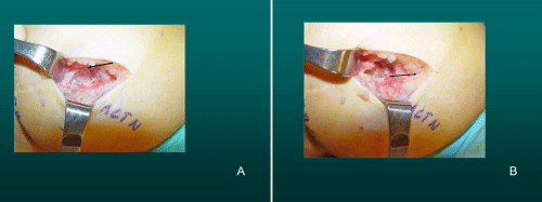
| Figure 7: Medial Knee Denervation. A) Incision is shown, medial to patella, centered over the tender site shown in Figure 3. The medial retinaculum is opened. The arrow points to the medial retinacular nerve adjacent to the recurrent medial recurrent geniculate vessels. B) The nerve has been resected. The arrow points to the direction in which, beneath the medial retinaculum, the nerve has been implanted into the vastus medialis muscle. C) Closure of the medial retinaculum is shown. |