|
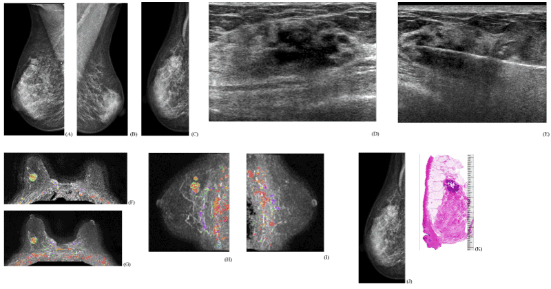
| Figure 2: Circular/oval-shaped invasive lobular carcinoma. This 83 year old woman felt a hard lump in the upper-outer quadrant of her right breast. Clinical breast examination confirmed the presence of a hard, clinically malignant tumor, 5 cm deep to the right nipple at the 11 o’clock position. Figures 2.A-C: Right and left mediolateral oblique projections. The palpable tumor is marked with a lead pellet in the upper portion of the right breast. The lateromedial horizontal projection demonstrates an ill-defined, circular lesion, partly hidden in the surrounding dense breast tissue. Figures 2.D-E: Hand-held ultrasound: a 22x11 mm hypoechoic, lobulated malignant tumor. Ultrasound guided 14-g core biopsy was performed. Histology: invasive lobular carcinoma, solid type. Figures 2.F-I: Breast MRI, left breast and axilla: no demonstrable abnormality; right breast, six cm deep to the nipple, in the upper half of the breast, there is a 17x15x16 mm round tumor with heterogeneous contrast enhancement. Both morphology and kinetics are characteristic for malignancy. An additional malignant tumor focus is seen 5 mm proximal to the larger tumor. Total disease extent: 17x15x28 mm. One lymph node in the right axilla is suspicious for metastases. Figures 2.J,K: Comparison of the lateromedial horizontal projection with large section histology of the same region. Histology report: multifocal invasive lobular carcinoma. The larger focus is histologically a solid type invasive lobular cancer that measures 17x12 mm (shown on Figure 2J); the satellite focus is a classic invasive lobular cancer measuring 1x11 mm. Biomarkers: ER/PR +ve, Her-2 -ve, Ki67 2%. LCIS was seen on a region measuring 40x45 mm. No metastases were found in the four surgically removed lymph nodes (pN 0/4). |