|
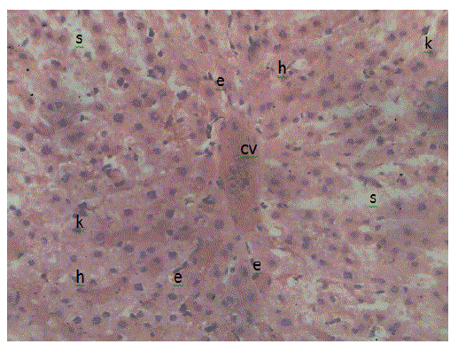
| Figure 5: Photomicrograph of the liver in the control group. Showing the hepatocytes arranged in plates radiating outwards from the central vein (cv), separated by sinusoids (s). Hepatocytes (h) are seen as polygonal shaped cells with large one or two centrally placed nucleus. Sinusoids are lined by flattened endothelial cells (e) continuous with that of the central vein. Resident macrophages (kuppfer cells, k) are seen as large irregularly shaped cells within the sinusoids. Magnification (x25). |