|
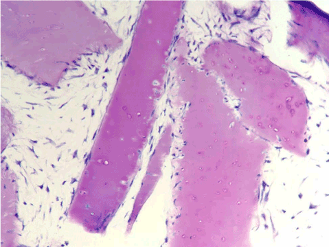
| Figure 4: Histological section of cartilage fluff matrix with cultured chondrocytes attached to the strands and growing in between the strands, after 40 days in culture. Cells penetrate and repopulate non- viable cartilage matrix. Romanowski-Giemsa stain. ×100. |