 |
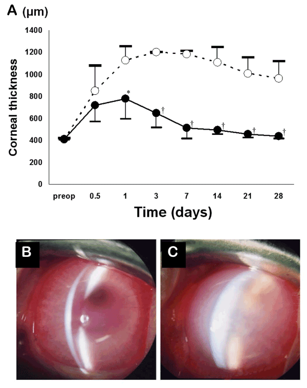
| Figure 4: Central corneal thickness in the control group (open circles) and the DSAEK group (closed circles) (A) and anterior segment photographs obtained with a slit-lamp microscope at 28 days after surgery (B, C). In the control group, the mean corneal thickness remains at around 1,000 μm for 28 days (A). In contrast, the mean corneal thickness gradually decreases in the DSAEK group and becomes significantly less than in the control group (A). There are significant differences of corneal thickness between the DSAEK and control groups on days 1, 3, 7, 14, 21, and 28. *p<0.05, †p<0.01 (A). (B) Representative anterior segment from the DSAEK group, showing a thin cornea without stromal edema. The margin of the pupil is clearly observed. (C) Severe corneal edema is observed in the control group. Details of structures in the anterior chamber cannot be visualized (C). |