|
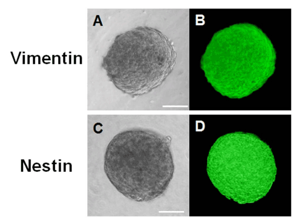
| Figure 8: Immunocytochemistry of sphere colonies derived from cultured HCECs on day 7. Bright field images (A, C) and immunostaining (B, D) of spheres are shown. The spheres were stained for vimentin (a mesenchymal cell marker) and nestin (a neural stem cell marker). On day 7, a sphere is nestin and vimentin-positive. Scale bar=100 μm. |