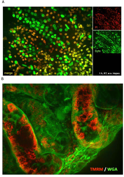
(A) A combination of PI and Syto 16 was used to visualize the nuclei of dead (PI,
red) and the nuclei of all cells (Syto 16, green) in the tubular area of the kidney.
The insets at the top right side shows the single channels, the big image shows
the merged image. The biopsy was incubated for one hour in standard culture
medium at RT. Note the heterogeneity in cell death between neighboring tubules.
(B) This image shows the result after staining such a kidney biopsy with
TMRM (red) and WGA (green). Like in (A) the heterogeneity in cell survival
is documented by TMRM fluorescence. Images were acquired using a 40x
water immersion objective.