|
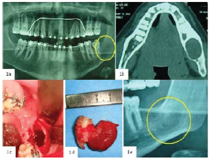
| Figure 1: Case 1: (a) OPT shows a unilocular radiolucent lesion in the molar region of the left mandible involving the roots of a permanent tooth. (b) Computed tomography confirms the presence of an osteolytic lesion in the posterior area of the mandible which caused the cortical bone expansion. (c) Intra-operatory view of the cavity after lesion removal and osteoplasty with piezoelectric ultrasonic tools. (d) Surgical specimen associated with a permanent tooth which has been extracted because of the association of its roots with the tumour. (e) OPT highlights the complete healing of the lesion after 1 year. |