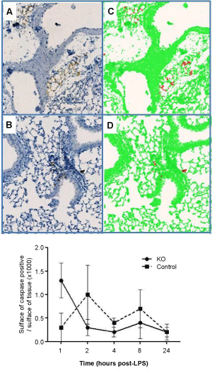
 Lung sections from IKK-2 (A) and littermate controls (B) twenty four hours
after exposure to LPS were stained for active caspase-3 and area positively
stained quantified by Definiens image analysis software (C and D). Images of
×20 magnification are shown. The geometric mean of the ratio of positively:
negatively stained areas (±SEM) for both groups at each time point is shown (E)
Lung sections from IKK-2 (A) and littermate controls (B) twenty four hours
after exposure to LPS were stained for active caspase-3 and area positively
stained quantified by Definiens image analysis software (C and D). Images of
×20 magnification are shown. The geometric mean of the ratio of positively:
negatively stained areas (±SEM) for both groups at each time point is shown (E)