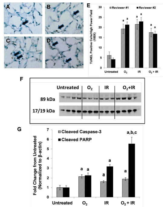
Panels A-D Histological lung sections of representative animals from each cohort (O2, IR, O2+IR) processed for TUNEL positivity (late phase, 20 weeks). Negative controls (performed simultaneously but without TdT) were examined at the same time. Panel E Cells were counted as % positive cells per high power field (400X) in 10 fields per condition. Data is represented as mean ± SEM. p< 0.05 for each challenge condition cohort vs. untreated as determined by two independent reviewers. Panel F Western blot for cleaved caspase 3 and cleaved PARP. Panel G Densitometric analysis of representative blot. Data is represented as mean fold change from untreated at each respective time point ± SEM. Letter a indicates p< 0.05 for all challenges vs. untreated at each respective time point. Letter b indicates p< 0.05 for O2+IR vs. O2 at each respective time point. Letter c indicates p< 0.05 for O2+IR vs IR at each respective time point.