 |
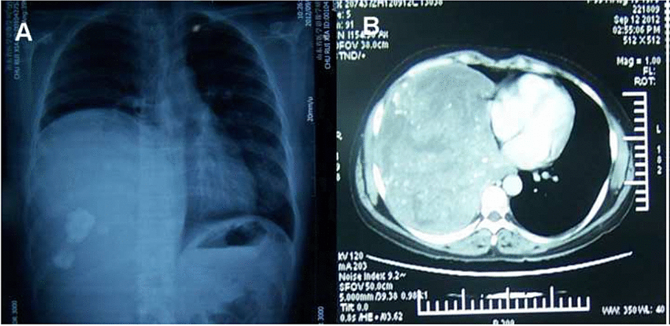
| Figure 1: Chest radiograph of a 33-year-old female patient showed solid mass within the right hemithorax (A). Chest computed tomography reveals a huge solid mass containing foci of intralesional calcifications in the right lower lobe of the lung (B). |