|
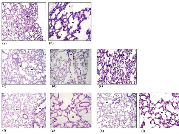
| Figure 11: Histopathological images representing the infiltration of inflammatory cells and RBCs in lung of rats exposed to hypobaric hypoxia at 7620 m, for 6 h. (a) Low power photomicrograph (10X) of lung parenchyma of normoxia animal showing a terminal bronchiole (TB) and alveolar spaces (AS). (b) High power (40X) photomicrograph from the same section of lung showing the clear alveolar spaces (AS) with thin septae; (c). Low power (10X) photomicrograph of patchy area of collapse of lung parenchyma of hypoxia exposed animal. The alveolar spaces (AS) are occluded by the collapsed septae (arrow). (d). Low power (10X) photomicrograph of another area of lung parenchyma of hypoxia animal showing an emphysematous pattern. (e). High power (40X) photomicrograph of lung of hypoxia animal from the collapsed area showing alveolar spaces (AS) with thickened septae (S) and inflammatory cell infiltration (ICs). Red blood cells (RBC) are seen within some of the alveolar spaces; (f) Low power (10X) photomicrograph of normoxia animals administered with curcumin showing lung parenchyma with a medium bronchus (BL) and alveolar spaces (AS). (g). High power (40X) photomicrograph from the same section of lung showing the clear alveolar spaces (AS) with thin septae; (h). Low power photomicrograph of lung parenchyma of hypoxia animals administered with curcumin, showing a terminal bronchiole (TB) and alveolar spaces (AS). No area of collapse or emphysema was seen. (i). High power (40X) photomicrograph from the same section of lung showing the clear alveolar spaces (AS) with mild thickening of some septae with no inflammatory cells. |