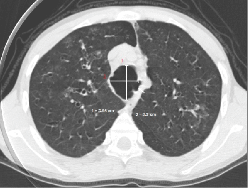
Figure 2:
High resolution computed tomography of chest shows abnormal dilatation of trachea in both transverse and anteroposterior diameter. Diverticulae are also seen (marked by arrow).