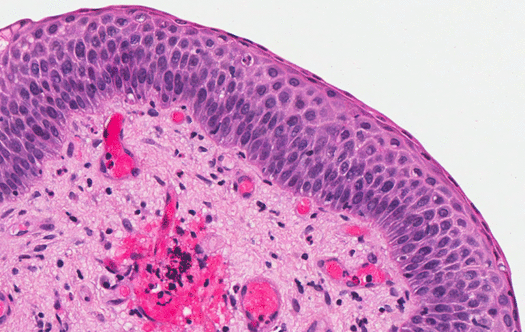
Figure 2c:
High magnification of the squamous component shows flattened surface epithelial cells without atypia (hematoxylin and eosin stain, original magnification x 40).