|
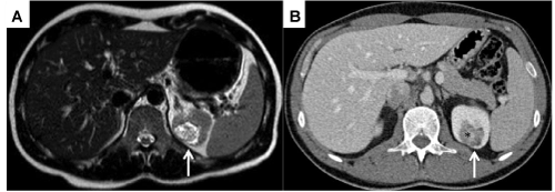
| Figure 1: A-B Incidental left renal mass. A, axial MRI T2 image showing iperintense area (arrow) in the upper portion of left kidney. B, axial post-contrast CT image showing low density area (arrow) in the upper portion of left kidney, with central contrast enhancement (asterisk). |