|
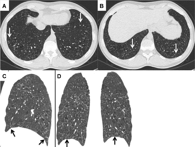
| Figure 2: A-D: A-B, axial CT image at the level of costo-phrenic sulci. Bilateral cysts are seen (arrows) involving costo-phrenic sulci down to their deeper portion. The cysts present homogeneous size and shape. C-D, sagittal (C) and coronal (D) CT image allow panoramic view of pulmonary cysts distribution. Involvement of lower lobes including costo-phrenic sulci (arrows) is shown in association with relative sparing of upper lobes. |