|
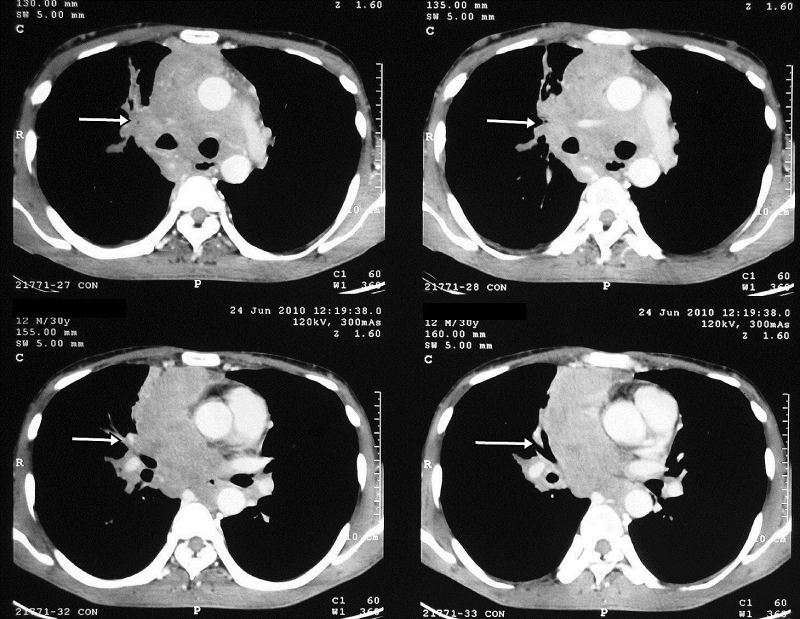
| Figure 2: CT Scan chest revealed a soft tissue infiltrative mass in anterior, superior and posterior mediastinal compartment with compression and displacement of superior vena cava and arch of aorta. Bilateral Hilar Lymph adenopathy with few nodular lesion in upper and middle lobe of right lung with involvement of pericardium. |