 |
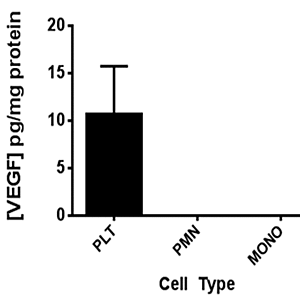
| Figure 2: VEGF content within pellets of apheresis platelets (PLT), whole blood granulocytes (PMN), and whole blood monocytes (MONO) normalized to mg total protein, expressed as mean ± SEM. At monocyte and granulocyte concentrations 100-fold greater (2 x 10⁴ cells/ml) than the small numbers of these cells contaminating platelet units, no detectable VEGF was found in lysates. Platelet lysates (1011 cells) had substantial VEGF content. Results are depicted as group mean ± SEM |