|
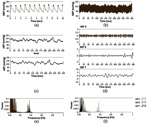
| Figure 2: The illustration of spectral analysis of BPV, including (a) The ABP signal with systolic blood pressure (sBP) peak detection (solid dot) within 10 seconds (b) The ABP signal within 300 seconds (c) top graph showed the systogram and bottom graph showed sBP time series after interpolation (d) IMF5-7 of ABP signal (e) FFT Spectrum of sBP time series (f) FFT Spectrums of IMF5-7. |