|
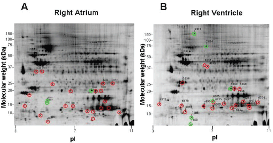
| Figure 2:Two-dimensional electrophoresis gels for the right atrium (A) and right ventricle (B). Twenty-four spots were chosen for subsequent mass spectrometry from each gel, identified by a green circle (greater than 1.5 fold increase, DMCT versus Sham) or red circle (greater than 1.5 fold decrease, DMCT versus Sham). |