|
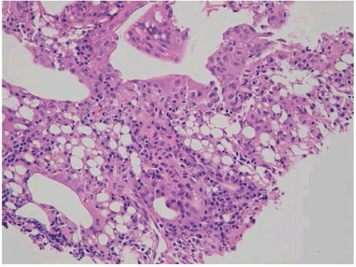
| Figure 2: Histological examination of TBLB showed fill-in alveoli by a large cluster of foamy lipid-filled macrophages, interstitial fibrosis around lipoid vacuoles, inflammatory lymphocytic infiltrates, some multinucleated foreignbody cells and fibrosis separating large vacuoles. |