 |
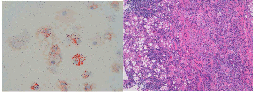
| Figure 4: (a) Cytological Oil Red O stain of BAL fluid showing the lipid nature of the vacuoles (red stained). (b) The histological examination of TBLB shows variable sized fat droplets, inflammatory lymphocytic infiltrates, interstitial fibrosis around lipoid vacuoles, and a large cluster of foamy lipid-filled alveolar macrophages. |