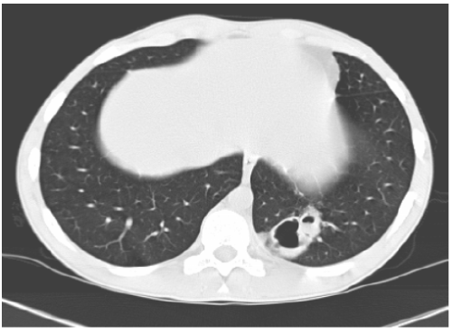
Figure 6:
Thin-walled pneumatoceles on the right lower lobe, with a mild perilesional ground glass attenuation. The surrounding parenchyma was normal.around lipoid vacuoles, and a large cluster of foamy lipid-filled alveolar macrophages.