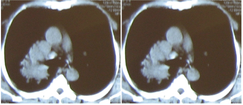
Figure 2:
Contrast enhanced computerized tomography of the thorax showing soft tissue density mass with irregular margins in the right upper lobe with obliteration of the lumen of right upper lobe bronchus.