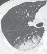
Figure 1:
Streptococcus pneumoniae
pneumonia. Computed tomography scan of right lobe showing airspace consolidation and ground glass opacities with a peripheral non-segmental distribution.