|
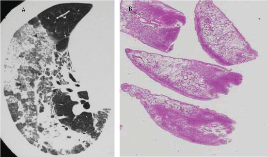
| Figure 10: COP. Computed tomography scan (A) is of the of right lower lobe shows diffuse airspace consolidation and ground-glass opacification with a peripheral non-segmental distribution, features suggesting COP. Lung biopsy specimen (B) displaysCOP with organization of inflammatory exudates in respiratory bronchiolar airspaces near the pleura. The alveoli far from the inflammatory bronchioles are relatively normal. |