 |
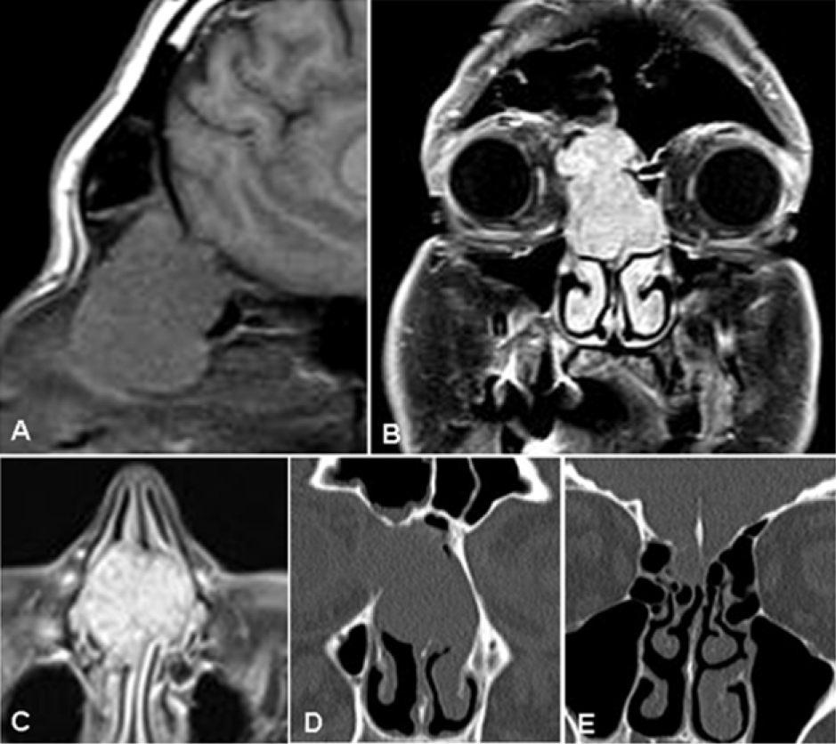
| Figure 1: MR/CT diagnostic work-up. Solid mass showed hypointense signal on sagittal T1 weighted images (A) with net and homogeneous enhancement after intravenous administration of gadolinium contrast agent on coronal (B) and axial (C) planes. CT images (D and E) demonstrated bone remodelling of the cribriform lamina and the medial wall of the right orbit. |