 |
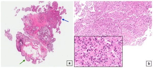
| Figure 2: A: Whole tissue mount of the sphenoid mass biopsy (10x magnification) showing normal olfactory epithelium at one end (green arrow) and the tumor mass at the other end (blue arrow), and B: same tumor mass at 100X magnification showing a vague hepatoid architecture (inset: same tumor at higher magnification of 400X). |