|
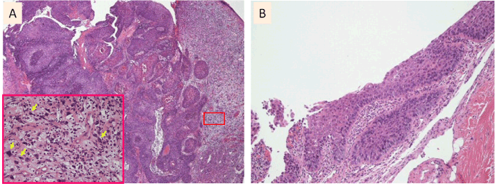
| Figure 4: Pathological examination before (A) and after (B) induction chemotherapy involving the administration of S-1 for two weeks followed by surgery. A: The tumor displayed a biphasic appearance involving the coexistence of a spindle cell component (right side) and a squamous cell carcinoma component (left side). The inset shows a magnified image of the pleomorphic spindle cells (arrow) in the boxed region. B: A pathological examination of the surgical specimen revealed carcinoma-in-situ without invasion into the surrounding tissues. |