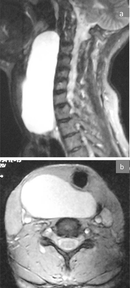
Figure 1: A, B:
Sagittal and axial (T1) MRI scans demonstrating a hyperdense neck mass located in the retropharyngeal space and having a mass effect on the trachea which is deviated to the left.