|
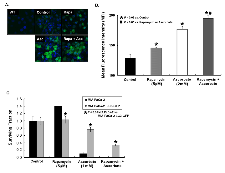
| Figure 2: Rapamycin enhances ascorbate-induced cytotoxicity despite increases in LC3-II punctae. A) MIA PaCa-2 cells (wild type or WT) and MIA PaCa-2 LC3-GFP cells were treated with the inducer of autophagy, rapamycin (5 μM) for 24 h, ascorbate (2 mM) for 1 h, or rapamycin followed by ascorbate. Cells were prepared for confocal microscopy using TO-PRO-3 as a nuclear stain and their inherent GFP expression as a marker for LC3 localization. As expected, wild type cells demonstrated no GFP fluorescence. Untreated MIA PaCa-2 LC3-GFP cells (Control) demonstrated a diffuse cytoplasmic GFP fluorescence with minimal appearance of GFP punctae. Treatment with rapamycin (Rapa) or ascorbate (Asc) led to a greater amount of GFP punctae compared to control, and treatment with the combination (Rapa+Asc) led to an even greater degree of punctae formation. B) Additional cells from the treatment groups in Figure 2A were prepared for flow cytometry to quantify the mean fluorescence intensity (MFI) of GFP in response to treatment. Cells were harvested and resuspended in propidium iodide (1 μg/mL) to sort for cell viability. Cells were gated for viability (PI negative) and GFP positivity, and MFI was quantified. Both rapamycin and ascorbate alone treatments had significantly greater MFI compared to untreated MIA-PaCa-2 LC3-GFP cells (146 ± 3 and 177 ± 4 respectively vs. 129 ± 1, P<0.05). Additionally, the combination of rapamycin + ascorbate induced an even greater increase in MFI compared to either treatment alone (195 ± 2, P<0.05). C) MIA PaCa-2 and MIA-PaCa-2 LC3-GFP cells were treated with rapamycin (5 μM) for 24 h, ascorbate (2 mM) for 1 h, or rapamycin followed by ascorbate. Cells were washed, trypsinized, and plated for clonogenic survival assay. Colonies were allowed to form for 10 days, and plates were fixed and stained with Coomassie blue. Colonies containing greater than 50 cells were scored. Surviving fraction was significantly decreased in both cell types when rapamycin was combined with ascorbate compared to either treatment alone. However, overall LC3 overexpressors exhibited enhanced clonogenic survival compared to their parent cellsafter both autophagy-inducing treatments (0.34 ± 0.02 for MIA PaCa-2 LC3-GFP vs. 0.01 ± 0.01 for MIA PaCa-2 after rapamycin+ascorbate, P<0.05). |