 |
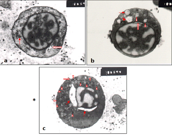
| Figure 1: (a, b, c). TEM. Peripheral blood withdrawn from mothers having autistic children. a) Showing, plasma cell with centric large sized nucleus, radially arranged dense clumps of heterochromatin, distinct perinuclear area of the cytoplasm, dilated Golgi cisternae (bold arrow), large number of various sized vesicles; thin arrow: pointed at large vesicle containing granules. b) Showing, altered plasma cell nucleus, the heterochromatin shifted to inside instead of being arranged directly underneath the nuclear membrane, nuclear pocket type I (bold arrow). The cytoplasm houses large number of ribosomes, loss of mitochondrial ultrastructure (arrow head), ultivescicular body (bent arrow) and autophagosome (double arrow), c) Showing, plasma cell with ruptured plasma membrane (bold arrow), with altered nucleus, highly dilated perivesceral nuclear space (thin arrow), the cytoplasm possesses numerous mitochondria and small sized dense granules (arrow head). [Specimen fixed in 4F1G and double-stained with Uranyl acetate and Lead citrate.X, 10000, 10000, 7500]. |