|
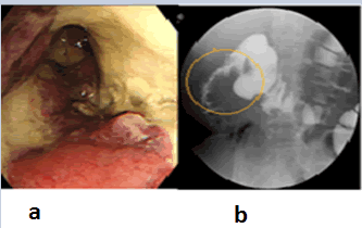
| Figure 2: (a) Colonoscopy revealed a circumferential white coat and necrotizing mucosa. (b) Gastrografin enema during colonoscopy revealed extensive stenotic lesions in the right-sided colon. (c) Microscopic specimens revealed marked infiltration of inflammatory cells. |