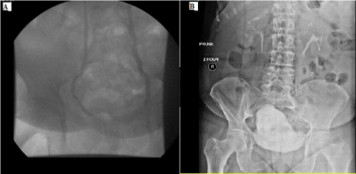
Figure 1:
A) Preoperative antegrade pyelography showing a lack of continuity of the distal ureter. B) Postperative Intravenous pyelography with normal upper urinary tract and passage of contrast material to the bladder.