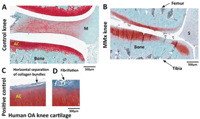
| Figure 7: Safranin O staining in the articular cartilage. Control (A), and MMx (B) knee joint. Human OA knee articular cartilage, served as positive control (C, D).
Articular cartilage from MMx knee as well as from control knee stained with Safranin O showing red coloration (8A, 8B). However, note the substantial loss of staining,
dominantly in the medial aspect of tibia including fibrillation area (white arrowheads), and in the femur of MMx knee (8B). Articular cartilage from human OA knee
shows positive staining with safranin O. However, there was a loss of staining in the region of longitudinal separation of collagen bundles (C) and fibrillation (D). |

